BIO 136
Human Anatomy and Physiology for Non-Majors
Study Guide
|
The Nervous System |
| Functions of the nervous system:
1) Integration of body processes 2) Control of voluntary effectors (skeletal muscles), and mediation of voluntary reflexes. 3) Control of involuntary effectors ( smooth muscle, cardiac muscle, glands) and mediation of autonomic reflexes (heart rate, blood pressure, glandular secretion, etc.) 4) Response to stimuli 5) Responsible for conscious thought and perception, emotions, personality, the mind. Structural Divisions of the nervous system: 1) Central Nervous System (CNS) - the brain and spinal cord. 2) Peripheral Nervous System (PNS) - the nerves, ganglia, receptors, etc. Functional Divisions of the Nervous System: 1) The Voluntary Nervous System - (a.k.a. somatic division) control of willful control of effectors (skeletal muscles) and conscious perception. Mediates voluntary reflexes. 2) The Autonomic Nervous System - control of autonomic effectors - smooth muscles, cardiac muscle, glands. Responsible for "visceral" reflexes. |
| Nervous System Histology - cell types:
1) Neurons - the functional cells of the nervous system. See below. 2) Neuroglia (glial cells) - Long described as supporting cells of the nervous system, there is also a functional interdependence of neuroglial cells and neurons. [See Glioma Tumors] a) astrocytes - these cells anchor neurons to blood vessels, regulate the micro-environment of neurons, and regulate transport of nutrients and wastes to and from neurons. [See Blood-Brain Barrier] b) microglia - these cells are phagocytic to defend against pathogens. They may also monitor the condition of neurons. c) ependymal cells - these cells line the fluid-filled cavities of the brain and spinal cord. They play a role in production, transport, and circulation of the cerebrospinal fluid. d) oligodendrocyte - produce the myelin sheath in the CNS which insulates and protects axons. [Multiple Sclerosis article] e) Schwann cells - produce the myelin sheath in the PNS. The myelin sheath protects and insulates axons, maintains their micro-environment, and enables them to regenerate and re-establish connection with receptors or effectors. Enables a rapid form of impulse conduction, called saltatory conduction. f) satellite cells - surround cell bodies of neurons in ganglia. Their role is to maintain the micro-environment and provide insulation for the ganglion cells. |
|
Neuron Structure:
In order to connect to other cells, receptors, and effectors, neurons have cytoplasmic extensions which attach to an enlarged area known as the cell body or cyton. Within the cell body is the nucleus and the neuron's biosynthetic machinery, the rough endoplasmic reticulum and the Golgi bodies. These organelles are so highly concentrated they can be visualized with a light microscope when stained with a specific technique. Called Nissl substance after the scientist who invented the staining technique, they manufacture the neurotransmitters which the neuron must secrete in large quantities. The neurotransmitter molecules are transported to the axon terminus by microfilaments and microtubules. There are two basic types of cytoplasmic extensions: the dendrites and the axon. Dendrites are short branching processes which receive stimuli from receptors or other neurons. They can perform this function because they, like the exposed membrane of the cell body, possess ion gates which respond to stimulation by neurotransmitters. A neuron will usually have only one axon, although it may branch extensively. The axon has ion gates which produce and transmit an action potential, and therefore it carries an impulse to another neuron or effector. At the end of the axon, the axon terminus, the neurotransmitters are released into the synapse. |
| Types of neurons based on structure:
The neuron pictured in Figure 7.4 is a multipolar neuron because it has many poles or processes, the dendrites and the axon. Multipolar neurons are found as motor neurons and interneurons (see below). There are also bipolar neurons with two processes, a dendrite and an axon, and unipolar neurons, which have only one process, which is classified as an axon. Unipolar neurons are found as most of the body's sensory neurons. Their dendrites are the exposed branches connected to receptors, the axon carries the action potential in to the central nervous system. |
| Types of neurons based on function:
motor neurons - these carry a message to a muscle, gland, or other effector. They are said to be efferent, i.e. they carry the message away from the central nervous system. sensory neurons - these carry a message in to the CNS. They are afferent, i.e. going toward the brain or spinal cord. interneuron (a.k.a. association neuron, connecting neuron) - these neurons connect one neuron with another. For example in many reflexes interneurons connect the sensory neurons with the motor neurons. |
| A simple Reflex Arc illustrates how these three types of neurons might work together. We will discuss types of reflexes in a later section. |
|
Spinal Cord and Peripheral Nerves |
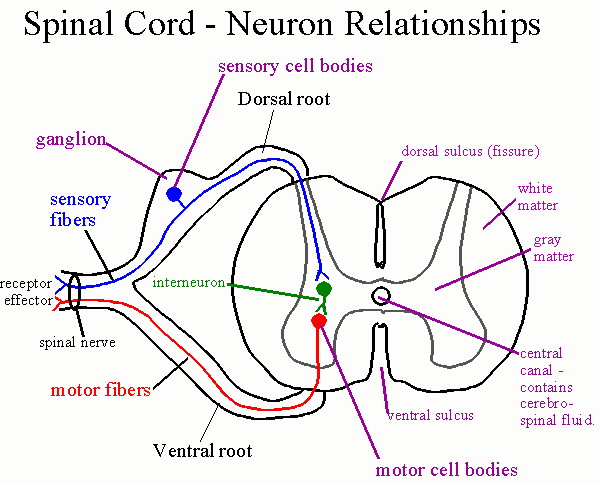
| The spinal cord is the connection center for the reflexes as well as
the afferent (sensory) and efferent (motor) pathways for most of the
body below the head and neck. The spinal cord begins at the
brainstem and ends at about the second lumbar vertebra. The
sensory, motor, and interneurons discussed previously are found in
specific parts of the spinal cord and nearby structures. Sensory
neurons have their cell bodies in the spinal (dorsal root) ganglion.
Their axons travel through the dorsal root into the gray matter of
the cord. Within the gray matter are interneurons with which the
sensory neurons may connect. Also located in the gray matter are
the motor neurons whose axons travel out of the cord through the
ventral root. The white matter surrounds the gray matter. It
contains the spinal tracts which ascend and descend the spinal cord.
Surrounding both the spinal cord and the brain are the meninges, a
three layered covering of connective tissue. The dura mater is the
tough outer layer. Beneath the dura is the arachnoid which is like a
spider web in consistency. The arachnoid has abundant space within
and beneath its thickened outer portion (the subarachnoid space) which contains
cerebrospinal fluid, as does the space beneath the dura mater
(subdural space). This cerebrospinal fluid supplies buoyancy for
the spinal cord and brain to help provide shock absorption. The pia
mater is a very thin layer which adheres tightly to the surface of the
brain and spinal cord. It follows all contours and fissures (sulci) of
the brain and cord.
An epidural injection of anesthetic, in childbirth for example, is placed immediately outside the dura mater. It penetrates slowly into the nearby nerve roots. |
| Terms:
ganglion - a collection of cell bodies located outside the Central Nervous System. The spinal ganglia or dorsal root ganglia contain the cell bodies of sensory neurons entering the cord at that region. nerve - a group of fibers (axons) outside the CNS. The spinal nerves contain the fibers of the sensory and motor neurons. A nerve does not contain cell bodies. They are located in the ganglion (sensory) or in the gray matter (motor). tract - a group of fibers inside the CNS. The spinal tracts carry information up or down the spinal cord, to or from the brain. Tracts within the brain carry information from one place to another within the brain. Tracts are always part of white matter. gray matter - an area of unmyelinated neurons where cell bodies and synapses occur. In the spinal cord the synapses between sensory and motor and interneurons occurs in the gray matter. The cell bodies of the interneurons and motor neurons also are found in the gray matter. white matter - an area of myelinated fiber tracts. Myelination in the CNS differs from that in nerves. |
| At 31 places along the spinal cord the dorsal and ventral roots come
together to form spinal nerves. Spinal nerves contain both sensory
and motor fibers, as do most nerves. Spinal nerves are given
numbers which indicate the portion of the vertebral column in which
they arise. There are 8 cervical (C1-C8), 12 thoracics (T1-T12), 5
lumbar (L1-L5), 5 sacral (S1-S5), and 1 coccygeal nerve. Nerve C1
arises between the cranium and atlas (1st cervical vertebra) and C8
arises between the 7th cervical and 1st thoracic vertebra. All the
others arise below the respective vertebra or former vertebra in the
case of the sacrum. Since the actual cord ends at the second lumbar
vertebra, the later roots arise close together on the cord and travel
downward to exit at the appropriate point. These nerve roots are
called the cauda equina because of their resemblance to a horses
tail.
Removing cerebrospinal fluid can be done by placing the needle in the sac of meninges below the conus medullaris where risk is minimal |
| The dermatomes are somatic areas served by fibers from specific spinal nerves. The map of the dermatomes is shown by. This map is useful in diagnosing the origin of certain somatic pain, numbness, tingling etc. when these symptoms are caused by pressure or inflammation of the spinal cord or nerve roots. Referred pain is caused when the sensory fibers from an internal organ enter the spinal cord in the same root as fibers from a dermatome. The brain is poor at interpreting visceral pain and instead interprets it as pain from the somatic area of the dermatome. So pain in the heart is often interpreted as pain in the left arm or shoulder, pain in the diaphragm is interpreted as along the left clavicle and neck, and the "stitch in your side" you sometimes feel when running is pain in the liver as its vessels vasoconstrict. |
| Spinal nerves join together in plexuses. A plexus
is an interconnection of fibers which form new combinations as the named peripheral nerves. There are
four voluntary plexuses (there are some autonomic plexuses which will be mentioned
later): they are the cervical plexus, the brachial plexus, the lumbar plexus, and the
sacral plexus. Each plexus gives rise to new combinations of fibers as the peripheral
nerves. The nerves and plexuses you need to know are:
Cervical Plexus - the phrenic nerve travels through the thorax to innervate the diaphragm. Brachial Plexus -
Lumbar Plexus
Sacral Plexus
|
| Structure of a
nerve:
A peripheral nerve is arranged much like a muscle in terms of its connective tissue. It has an outer covering which forms a sheath around the nerve, called the epineurium. Often a nerve will run together with an artery and vein and their connective coverings will merge. Nerve fibers, which are axons, organize into bundles known as fascicles with each fascicle surrounded by the perineurium. Between individual nerve fibers is an inner layer of endoneurium. The myelin sheath in peripheral nerves consists of Schwann cells wrapped in many layers around the axon fibers. Not all fibers in a nerve will be myelinated, but most of the voluntary fibers are. The Schwann cells are portrayed as arranged along the axon like sausages on a string. (A more apt analogy would be like jelly rolls!) Gaps between the Schwann cells are called nodes of Ranvier. These nodes permit an impulse to travel faster because it doesn't need to depolarize each area of a membrane, just the nodes. This type of conduction is called saltatory conduction and means that impulses will travel faster in myelinated fibers than in unmyelinated ones. The myelin sheath does several things: 1) It provides insulation to help prevent short circuiting between fibers. 2) The myelin sheath provides for faster conduction. 3) The myelin sheath provides for the possibility of repair of peripheral nerve fibers. Schwann cells help to maintain the micro-environments of the axons and their tunnel (the neurilemma tunnel) permits re-connection with an effector or receptor. (See below) CNS fibers, not having the same type of myelination accumulate scar tissue after damage, which prevents regeneration. See [Spinal Cord Repair] |
| Regeneration of a peripheral nerve fiber depends upon several things. First the damage must be far from the cell body. Anterograde degeneration destroys the axon distal to the point of damage. Retrograde degeneration causes the fiber to degenerate for a distance back toward the cell body. The amount of axoplasm lost determines whether the neuron can survive. Secondly the myelin sheath and its neurilemma tunnel must be intact. Chemicals such as the myelin proteins tend to inhibit regrowth, but macrophages will enter the damaged area and phagocytize these proteins and other debris. Schwann cells will proliferate and secrete growth stimulating factors and provide the chemical and physical needs necessary for growth and re-innervation by the axon. |
| The Spinal Tracts:
The white matter of the spinal cord contains tracts which travel up and down the cord. Many of these tracts travel to and from the brain to provide sensory input to the brain, or bring motor stimuli from the brain to control effectors. Ascending tracts, those which travel toward the brain are sensory, descending tracts are motor. Figure 12.30 shows the location of the major tracts in the spinal cord. For most the name will indicate if it is a motor or sensory tract. Most sensory tracts names begin with spino, indicating origin in the spinal cord, and their name will end with the part of the brain where the tract leads. For example the spinothalamic tract travels from the spinal cord to the thalamus. Tracts whose names begin with a part of the brain are motor. For example the corticospinal tract begins with fibers leaving the cerebral cortex and travels down toward motor neurons in the cord. [See Spinal Tract Pathways] White matter fibers are surrounded by oligodendrocytes which form the myelin sheath in CNS fibers. Diseases which destroy the myelin sheath lead to inability to control muscles, perceive stimuli etc. One such disease is multiple sclerosis, an autoimmune disorder in which your own lymphocytes attack the myelin proteins. [See Beta Interferon and Multiple Sclerosis]. Here are the major spinal tracts and their functions:
|
|
The Brain | Laberal Brain diagram | Sagittal Brain diagram |
| The brain develops from the anterior end of the neural tube. The central cavity of the tube is seen as the central canal of the spinal cord, and as cavities of the brain called ventricles which contain cerebrospinal fluid. The brain develops along a linear arrangement of structures, but due to their confinement in the cranium the linear nature of this arrangement is contorted. |
| Arrangement of gray and white matter:
In the spinal cord the gray matter is found only as an H-shaped area in the center of the cord. But within the brainstem the gray matter diverges, passing through the center of the cerebrum and terminating in the gray matter of the cortex. The cerebellum has its own gray and white matter distribution which is like that of the cortex. Other parts such as the basal nuclei have both gray and white matter. Gray matter in the brain functions much like that in the cord: it is the site of connections between neurons and contains the cell bodies of motor and interneurons. It is composed of unmyelinated neurons. White matter in the brain, like that in the spinal cord, is composed of myelinated fibers in tracts which carry information from one place to another. The ventricles: Ventricles are cavities within the brain derived from the original lumen of the neural tube. They exist in each cerebral hemisphere (the lateral ventricles), between the hemispheres (the third ventricle), and beside the cerebellum (the fourth ventricle). Canals connect these ventricles together and lead to the spaces of the meninges. |
| Circulation of the cerebrospinal fluid:
The cerebrospinal fluid (CSF) is produced by filtration from blood capillaries located in tissue known as the choroid plexus which lines the ventricles. Filtration forces water, electrolytes, and nutrients out of the blood. These substances are then processed by the ependymal cells which release them into the cerebrospinal fluid. At the same time waste products are removed from the CSF and released into the blood. The cerebrospinal fluid circulates between the ventricles and into the spinal canal. It also enters the subarachnoid and subdural spaces through apertures near the fourth ventricle (see Figure 12.24). It therefore also bathes the brain and spinal cord providing buoyancy. Eventually it must be reabsorbed into the bloodstream and this occurs at pockets of arachnoid tissue which invaginate into a large vein called the superior sagittal sinus. These pockets are called the arachnoid granulations or arachnoid villi and allow the fluid to move into the veins by osmosis and a pressure gradient produced by lower pressure in the vein. The absorption must occur at the same rate as production of CSF in order to prevent an imbalance. If absorption is insufficient it causes a condition known as hydrocephaly, "water on the brain". This condition usually shows up in early childhood and can damage the brain and lead to abnormal development. It is usually corrected surgically with a shunt, a small tube which drains fluid from the meninges and short circuits it into a nearby vein. |
| The Cerebral Hemispheres: The cerebrum is divided into two hemispheres separated by the longitudinal fissure. The terms
fissure and sulcus describe grooves in the surface of the cerebrum. Most of the time fissure refers to
a larger groove than a sulcus, although they are somewhat interchangeable. In between grooves is
found a raised area called a gyrus. Certain of these structures are consistent landmarks for the brain.
The outer layer of the cerebrum is composed of gray matter and is called the cerebral cortex. The
cerebral cortex is the area of conscious thought and perception. For this reason, and because it
forms a cap over the rest of the brain the cerebral cortex has been called the "thinking cap". The
cortex can be described as made up of regions called lobes. Each lobe bears the name of the bone
lying above it. The central sulcus separates the parietal from the frontal lobe. The lateral fissure
divides the parietal from temporal lobe. A short parieto-occipital fissure indicates the upper
delineation of the parietal from occipital lobe. The largest fissure is the longitudinal fissure which
divides the two hemispheres from one another.
The cerebral hemispheres are connected by fiber tracts which permit the hemispheres to communicate with one another. The major connection is the corpus callosum, a second is the fornix. The hemispheres normally divide up the tasks with one hemisphere, usually the left, being dominant. This is the principle of lateralization and dominance. In most people (90 to 95%) the left hemisphere is dominant and responsible for logic, mathematics, and language. The right hemisphere is the center for emotions and artistic endeavors. However, when one hemisphere is damaged the other may be capable of performing the lost functions. |
| The Projection Areas (Functional Areas) of the Cerebrum:
Projection areas are regions of the cortex where specific motor or sensory activity is localized. The pre-central gyrus (the raised area in front of the central sulcus) is the primary motor area. It is the center for voluntary control of the skeletal muscles. Each area of the body, for that matter each muscle, is controlled by a specific part of the pre-central gyrus. The orientation is generally reversed in position compared to the body location, i.e. the muscles of the leg and foot are generally projected to the top of the gyrus, those in the head and neck to the bottom of the gyrus. The area represented reflects the level of activity and control over the muscles, not their size. For example more area is devoted to the muscles of facial expression, speech, and hand movement as to all the rest of the muscles. The pre-central gyrus is the origin of the corticospinal tracts. The post-central gyrus is the comparable area for sensory perception, called the somatosensory area. It receives the conscious sensations from the musculocutaneous regions of the body: pain, temperature, touch, and pressure. These sensations are brought by the spinothalamic tract and the fasciculus gracilis and cuneatus. As before, these sensations terminate in specific positions on the gyrus which are inversely related to body position and directly related to the degree of sensation and its importance, not the size of the area. Other projection areas of importance: The pre-motor area - in front of the pre-central gyrus, a motor association area partially responsible for learned reflexes. Frontal eye field - synchronizes eye movements. Broca's area - the motor speech area for control of the muscles of speech. Prefrontal cortex - Important in planning complex movements and in general planning and elaboration of thoughts. This area normally exerts control over other areas such as those responsible for emotions and stress response, and is thus thought to be the center for self-control, reasoning, and such. It is responsible in part for personality and some aspects of memory. Wernicke's area - Language comprehension and elaboration; a general interpretive area. Primary auditory area - this area receives and perceives hearing. Primary visual area - this area perceives visual stimuli and constructs a three-dimensional image using stimuli from both eyes. |
| Other areas of the cerebrum:
The basal nuclei consist of a group of structures with both gray and white matter which in various ways modify motor functions coming from the cerebral cortex. The basal nuclei function in association with the corticospinal system to control complex patterns of motor activity. When there is serious damage the basal nuclei the motor cortex can no longer provide the patterns for many skilled and repetitive actions. Included are: writing letters of the alphabet, using scissors, hammering nails, shooting a basketball, passing a football, shoveling dirt, controlled eye movements and many others. Several circuits connect the basal nuclei with the motor association areas, sensory association areas, and the motor cortex in loops which provide both positive and negative feedback. The basal nuclei utilize a wide range of neurotransmitters: GABA (gamma amino butyric acid) and dopamine are inhibitory neurotransmitters. Ach, norepinephrine, serotonin, and enkephalin are found in connecting pathways, both excitatory and inhibitory, to other areas, and there are multiple glutamate pathways which provide most of the excitatory signals within the basal nuclei. Disorders:Parkinson's Disease - caused by destruction of dopamine secreting cells in the substantia nigra which send impulses to the caudate nucleus and putamen. The result is that without these inhibitory impulses, tremor and rigidity occur. Also akinesia occurs, or lack of the ability to perform willful movements. Treatments: 1) use of L-dopa which is converted into dopamine in the brain (eventually the brain resists this); 2) transplanted fetal dopamine cells and genetically engineered dopamine cells; 3) the use of the MAO inhibitor Deprenyl. MAO (mono amine oxidase) is the chemical which breaks down dopamine); growth factors which stimulate recovery or block deterioration of the dopamine cells. The activation of existing stem cells in the new frontier of research for possibly curing Parkinson's and other neurological disorders. [Parkinson's and Dopamine] [Parkinson's Article] [Interpreting Alzheimer's Abnormalities] [Alzheimer's Article] [The Prefrontal Lobes and Schizophrenia] [Schizophrenia Article] Huntington's Disease - Begins with flicking movements a joints which progress to severe distortional movements of the entire body leading eventually to severe dementia. It is caused by a genetic mutation of an enzyme-producing gene. This results in loss of GABA secreting neurons in the basal nuclei, and in loss of ACH neurons in many parts of the brain. Alzheimer's Disease - caused by the development of neurofibrillar tangles and beta-amyloid plaques in the brain. The tangles occur when a support protein called "tau", which is important in support of the transport proteins for the cell, breaks down, causing the proteins to intertwine and loose their functional transport structure. Without transport proteins neurons cannot function or survive. The plaques accumulate in large masses in certain people. In response to these the immune cells produce chemicals called cytokines which attack the plaques. Unfortunately these chemicals damage and kill the neurons they are supposed to protect. Treatments focus on various aspects of the problem. On one front factors which cause the breakdown of tau may be able to be blocked, thus preventing the tangles. On another front antibodies against the beta amyloid plaque have been developed which show promise in eliminating it in mice.
|
| Parts of the brain below the cerebrum:
[human brain sectioned] [Sagittal Section Diagram] corpus callosum - connects the hemispheres. The fornix - also connects hemispheres and part of the limbic system. septum pellucidum - a membrane which covers the opening into the lateral ventricle. The Dienchephalon The thalamus - its halves connected by the intermediate mass, the thalamus receives all conscious sensations and acts as a relay center. Sensations from the spinothalamic tract and the fasciculus gracilis and fasciculus cuneatus synapse in the thalamus before continuing to the cortex. Afferent impulses are routed by the thalamus to their proper destinations. The thalamus also lies at the top of the reticular formation and is part of the alert mechanism of the reticular activating system. The thalamus also helps to filter out unwanted stimuli. The hypothalamus is a small yet very important part of the brain below the thalamus (hypo=below). It is part of the control mechanism for many of the endocrine glands. Through its connection with the pituitary gland through the infundibulum the hypothalamus directs the pituitary's secretions, which in turn direct many other endocrine glands. The hypothalamus also coordinates many autonomic and visceral functions such as control of blood glucose, heart rate and respiration in response to stresses, control of thermoregulation, the perception of hunger, thirst, control of electrolyte and water balance, and the sleep-wake cycle. The pineal gland is considered part of the epithalamus (epi=upon the thalamus) and receives stimuli from the hypothalamus. The pineal gland secretes melatonin during the dark periods. This establishes our biological clock and regulates our circadian rhythm (day-night cycle) which affects many behaviors such as sleeping, eating, sexual desire, etc. Individuals who receive insufficient light, such as those living in the far north during the winter, may experience Seasonal Affective Disorder. This disorder affects their mood and mental state as well as their physiology. Lights which stimulate the retina in a pattern similar to that of normal sunlight has been shown to alleviate the problem. The Brainstem: Composed of the midbrain, pons, and medulla. The most important (to our study) part of the midbrain is the corporal quadrigemina. The corpora quadrigemina (four bodies, twins) are composed of the superior colliculi, which are the center for visual reflexes (blinking, accommodation of the lens), and the inferior colliculi where auditory reflexes are centered (contraction of the stapedius muscle). The pons - the name means bridge and in part the pons is a bridge to the higher brain. However its main functions are as part of the regulation of the rate and depth of respiration. It also contains the CNS connection for cranial nerves V, VI, VII, and VIII. The medulla - center for the vital functions of heart rate, respiration, and blood pressure. The medulla also has CNS connections for VIII, as well as IX, X, XI, and XII. The pyramids are also part of the medulla. The medulla stimulates the muscles of respiration and controls the breathing rhythm. It regulates the heart rate and volume and controls blood pressure and overall blood distribution. The cerebellum - this part coordinates the skeletal muscles. It receives unconscious proprioception as well as input from all the higher motor centers. From this input the cerebellum monitors muscle contractions and planned muscle contractions and maintains a constantly adapting system to coordinate them. |
| Functional (Conceptual) Areas of the brain: these areas are considered by some to work together to
perform certain functions, but are not related structurally.
Functions of the Limbic System - this system is sometimes called the "emotional brain". It is the site of: 1) emotional states and behavior. 2) the bridge between the conscious and subconscious brain. This is how subconscious feelings surface as voluntary behavior. 3) short term memory and information storage, especially short term recognition of facts, objects, people, etc. |
| The Reticular Formation and Reticular Activating System:
The Reticular Formation is a brainstem pathway which receives sensory input of many types including vision, auditory, and somatic senses. It directs these stimuli to the thalamus as part of the Reticular Activating System which is an alert system for make the cortex. This allows unwanted and unimportant stimuli to be filtered out, while making us aware of important and critical stimuli. |
| The Cranial Nerves |
Neurophysiology |
| This section deals with transmission of an action potential. This occurs in two ways:
1) across the synapse - synaptic transmission. This is a chemical process, the result of a chemical neurotransmitter. 2) along the axon - membrane transmission. This is the propagation of the action potential itself along the membrane of the axon. |
| Synaptic transmission - What you learned about the neuromuscular junction is mostly
applicable here as well. The major differences in our current discussion are:
1) Transmission across the synapse does not necessarily result in an action potential. Instead, small local potentials are produced which must add together in summation to produce an action potential. 2) Although Ach is a common neurotransmitter, there are many others and the exact effect at the synapse depends on the neurotransmitter involved. 3) Neurotransmitters can be excitatory or inhibitory. The result might be to turn off the next neuron rather than to produce an action potential. |
| The basic steps of synaptic transmission are the same as described at the neuromuscular
junction. The diagram looks very similar too. [See Synaptic Transmission]
But there are some important differences.
1) Impulse arrives at the axon terminus causing opening of Ca+2 channels and allows Ca+2to enter the axon. The calcium ions are in the extracellular fluid, pumped there much like sodium is pumped. Calcium is just an intermediate in both neuromuscular and synaptic transmission. 2) Ca+2 causes vesicles containing neurotransmitter to release the chemical into the synapse by exocytosis across the pre-synaptic membrane. 3) The neurotransmitter binds to the post-synaptic receptors. These receptors are linked to chemically gated ion channels and these channels may open or close as a result of binding to the receptors to cause a graded potential which can be either depolarization, or hyperpolarization depending on the transmitter. Depolarization results from opening of Na+ gates, hyperpolarization could result from opening of K+ gates. 4) Graded potentials spread and overlap and can summate to produce a threshold depolarization and an action potential when they stimulate voltage gated ion channels in the neuron's trigger region. 5) The neurotransmitter is broken down or removed from the synapse in order for the receptors to receive the next stimulus. As we learned there are enzymes for some neurotransmitters such as the Ach-E which breaks down acetylcholine. Monoamine oxidase (MAO) is an enzyme which breaks down the catecholamines (epinephrine, nor-epinephrine, dopamine) and nor-epinephrine (which is an important autonomic neurotransmitter) is removed by the axon as well in a process known as reuptake. Other transmitters may just diffuse away. |
|
Inhibition - When the brain causes a hyper-polarization in advance of a reflex pathway being stimulated, it reduces the likelihood of the reflex occurring by increasing the depolarization required. The pathway can still work, but only with more than the usual number or degree of stimulation. We inhibit reflexes when allowing ourselves to be given an injection or blood test for instance. Facilitation - When the brain causes a depolarization in advance of a reflex pathway being stimulated, it makes the reflex more likely to occur, requiring less additional stimulation. When we anticipate a stimulus we often facilitate the reflex. Learned Reflexes - Many athletic and other routine activities involve learned reflexes. These are reflex pathways facilitated by the brain. We learn the pathways by performing them over and over again and they become facilitated. This is how we can perfect our athletic performance, but only if we learn and practice them correctly. It is difficult to "unlearn" improper techniques once they are established reflexes. Like "riding a bike" they may stay with you for your entire life! |
| Comparison of the autonomic and somatic (voluntary) nervous
systems:
Both systems involve reflexes and connections to the CNS utilizing unipolar and multipolar sensory neurons, interneurons, and motor neurons. But there are some differences. Functional differences: The autonomic system is often considered a motor system for control of autonomic effectors. These effectors include smooth muscle, glands, and the heart. It also utilizes sensory inputs as part of visceral reflexes and independently as part of broader control mechanisms. Structural differences: While the motor neuron in a somatic pathway travels directly to the skeletal muscle effector, in the autonomic nervous system there are two motor neurons which synapse at an autonomic ganglion. The neurotransmitter also may vary with the parasympathetic division mostly using acetylcholine at both pre and post ganglionic fibers, while the sympathetic division utilizes mostly norepinephrine at the post ganglionic fibers. See below. |
| The Two Autonomic Divisions: Sympathetic and Parasympathetic
These two divisions innervate most of the same effectors, but their actions differ: See the PDF file, slide 76. The Parasympathetic Division - This division originates (has its CNS connection) in either the cranial or sacral portions of the CNS. That means it utilizes cranial nerves (remember III, VII, IX, X) or sacral spinal nerves (the pelvic splanchnic nerves). Its autonomic ganglia are located on or near the effector organ. It utilizes acetylcholine as both the pre-ganglionic and the post-ganglionic neurotransmitter ( we call that cholinergic). The post-ganglionic neurotransmitter is most important because this is the transmitter actually released at the effector organ and which stimulates its receptors. The parasympathetic division has localized and specific effects which can mostly be described as producing the "normal" responses, i.e. non-stress responses, of its effectors. The Sympathetic Division - This division has its origin in the thoracolumbar portion of the spinal cord. Its autonomic ganglia are located in a chain of ganglia (called the lateral chain ganglia) located near the spinal cord. There are also plexuses which allow multiple connections between different components of the division. The sympathetic division uses ACH at its autonomic ganglia, but mostly uses norepinephrine at the effector organ (adrenergic). This permits an entirely different effect on many of the same effectors as the parasympathetic division. In addition the sympathetic division often exhibits a general activation rather than local effect. The sympathetic division often acts as a "stress response" system. The alliteration "Fight or Flight" is sometimes used to describe the way in which the sympathetic division becomes activated to mobilize the body's resources. |
)
)
)
)
)
)
)
)
)
)